
Cytotoxic lesion of the corpus callosum in a patient with aphasic status epilepticus
##plugins.themes.bootstrap3.article.sidebar##
Published
Dec 7, 2020
##plugins.themes.bootstrap3.article.main##
Abstract
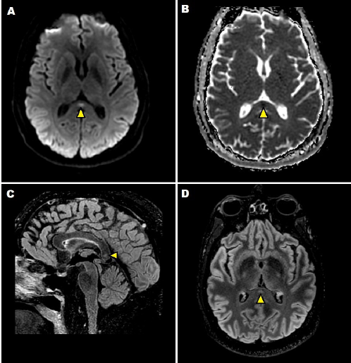
A 47-year-old man with a history of aphasic seizures presented to the emergency room with a 12-hour global aphasia. Upon admission, brain MRI did not reveal acute lesions, and EEG showed sharp waves in the left frontal-temporal region. An Aphasic Status Epilepticus was diagnosed and antiepileptic treatment was initiated with adequate response. A week after the episode, a new brain MRI showed a high-signal ovoid lesion on T2-weighted and FLAIR sequences in the central part of the splenium of the corpus callosum.
Downloads
Download data is not yet available.
##plugins.themes.bootstrap3.article.details##
How to Cite
Castiglione, J.-I., Ricciardi, M.-E., & Bensi, C. (2020). Cytotoxic lesion of the corpus callosum in a patient with aphasic status epilepticus. Journal of Applied Cognitive Neuroscience, 1(1), 98–100. https://doi.org/10.17981/JACN.1.1.2020.15
Section
Neuroimage

This work is licensed under a Creative Commons Attribution-NonCommercial-NoDerivatives 4.0 International License.
You are free to:
- Share — copy and redistribute the material in any medium or format.
- The licensor cannot revoke these freedoms as long as you follow the license terms.
Under the following terms:
- Attribution — You must give appropriate credit, provide a link to the license, and indicate if changes were made. You may do so in any reasonable manner, but not in any way that suggests the licensor endorses you or your use.
- NonCommercial — You may not use the material for commercial purposes.
- NoDerivatives — If you remix, transform, or build upon the material, you may not distribute the modified material.
- No additional restrictions — You may not apply legal terms or technological measures that legally restrict others from doing anything the license permits.